Multi-color luminescent screens for label-free bio-imaging
Référence
04585-01
Mots-clés
Statut des brevets
French priority patent application n°FR2981159 filed on october 11th, 2011 and entitled “Procédé de détection de l’interaction d’au moins une entité avec une couche diélectrique”


Inventeurs
Volodymyr LYSENKO
Tetyana NICHIPORUK
Tetiana SERDIUK
Yuri ZAKHARKO
Alain GEOLEN
Mustapha LEMITI
Statut commercial
Exclusive or non-exclusive license
Laboratoire
Nanotechnology Institute of Lyon (INL, UMR5270), Lyon, France
Description
CONTEXT
Multicolor imaging of bio-objects by one-photon excited fluorescence usually requires their labeling by various external molecular fluorophores, fluorescent proteins or quantum dots. While the major drawback of quantum dots is their cytotoxicity, organic fluorophores are strongly affected by photobleaching. Moreover, any external fluorescent agents affect the intracellular environment. Thus, developement of a label-free optical approache could be a powerful cell imaging tool. We propose to use silicon-based nanostructured dielectric thin films as luminescent screens for label-free bio-imaging
TECHNICAL DESCRIPTION
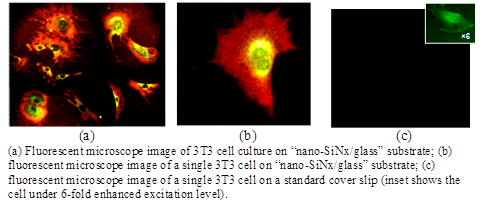
The nano-structured silicon-based fluorescent 40-100 nm thin films can be deposited on almost any suitable solid-state substrate. Cells which have to be observed are grown on the films without additional labelling. Specific physico-chemical interactions between different cell compartments and the luminescent screens on which the cells are immobilized, switch on various luminescent mechanisms in the substrates, ensuring multicolour imaging of the studied cells. Thus, subcellular components and/or organelles (nucleus, mitochondria, endoplasmic reticulum, cytoplasm …) can be easily visualized. Intensity of the cell coloration is much stronger than monocolour (green) auto-fluorescence signal of the same cells deposited directly onto the usual glass substrates (cover slips) and observed under the same conditions (excitation intensity and acquisition time).

DEVELOPMENT STAGE
Manufacturing of the «fluo-screen/glass» substrates is perfectly mastered by INL researchers. Current production rate is about 1200 substrates (1,5 cm x 1,5 cm) per hour. This new imaging approach was tested and validated in-vitro with various healthy and cancerous (animal and human) cell cultures.
BENEFITS
Our luminescent screens:
- allow label-free multicolour cell imaging;
- are totally compatible with standard fluorescent microscopy;
- are biocompatible and are friendly to the environment;
- may be disposable;
- have low production cost.
INDUSTRIAL APPLICATIONS
Accessories for bio Imaging systems
For further information, please contact us (Ref 04585-01)
Besoin de plus d'informations ?
Nous contacterTechnologies Liées
-
15.02.2017
Describe : Additive and subtractive submicro-manufacturing
Dispositifs & Instruments, Matériaux – Revêtements 05975-01
-
04.02.2014
Transparent glass and ceramic glass in a large wavelength bandwidth (from visible to infrared 6µm)
Matériaux – Revêtements 05728-01
-
04.10.2013
Self healing vitreous composition, their preparation and uses
Matériaux – Revêtements 02782-01